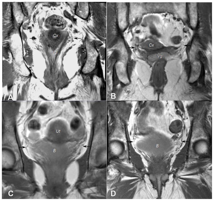
Figure 3.
Coronal scans from 4 different women illustrate attachments of the cardinal ligament to the pelvic organs. A, B, Black arrows point to the cardinal ligament’s insertions at both the cervix (Cx) and upper vagina (Vg). C,D, Fibers of the cardinal ligament, again identified with black arrows, continue to the bladder (B).

