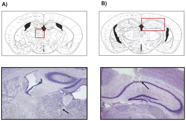
Figure 4.
Histological sections showing placement of stimulating and recording electrodes in the region of midline thalamic nuclei (A) and in the CA1 region of the hippocampus (B) respectively. Position of the electrode for electrical stimulation in the midline thalamic nuclei was −1.1 mm posterior to bregma and the position of the electrode for recording in the CA1 region of the hippocampus was −2.1 mm posterior to bregma.

