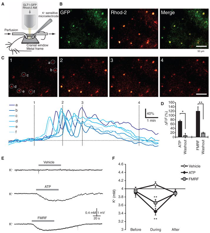
Fig. 8.
Agonist-induced increases in astrocytic Ca2+ reduce extracellular K+ concentration in vivo. (A) Experimental setup for in vivo recordings. (B) Glt1-eGFP reporter mouse loaded with the Ca2+ indicator rhod-2 AM. Rhod-2 only–labeled eGFP+ astrocytes. (C) Time-lapse images of ATP (100 μM)–evoked increases in Ca2+ in astrocytes located 95 μm below the pial surface. Colored traces indicate relative changes in rhod-2 signals of astrocytes (a to f) labeled in the first panel. (D) Comparison of ATP (100 μM)– and FMRF (15 μM)–induced increases in rhod-2 signal (ΔF/F). n = 4 to 5 mice. *P < 0.05; **P < 0.01, paired t test. (E) Representative recordings of extracellular K+ concentration in mouse cortex exposed to vehicle (aCSF), ATP (100 μM), or FMRF (15 μM). (F) Graph comparing the effects of vehicle, ATP, and FMRF on extracellular K+. All recordings were obtained 100 μm below the cortical surface. n = 7 mice. **P < 0.01, Bonferroni-Dunn test compared to baseline.

