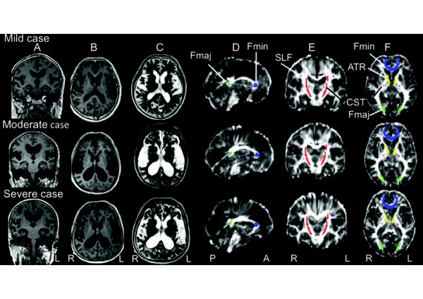
Figure 1.
Magnetic resonance images from three idiopathic normal pressure hydrocephalus (INPH) patients arranged in descending order of the anterior thalamic radiation fractional anisotropy (FA) level (mild, moderate and severe). A) T1-weighted image (coronal view). B) T1-weighted image (axial view). C) T2-weighted image (axial view). D) Diffusion tensor image FA map (sagittal view). E) Diffusion tensor image FA map (coronal view). F) Diffusion tensor image FA map (axial view). Regions of interest (ROI) colors: forceps minor (Fmin, blue); anterior thalamic radiation (ATR, yellow); superior longitudinal fasciculus (SLF, purple); corticospinal tract (CST, red); forceps major (Fmaj, green).

