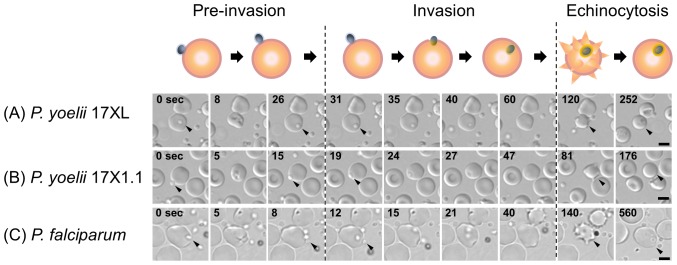
Figure 2. Three phase processes of the red blood cell (RBC) invasion by Plasmodium yoelii.
Time-lapse imaging of RBC invasion was captured every 0.1 sec with transmitted light for P. yoelii 17XL (A), P. yoelii 17X1.1 (B), and Plasmodium falciparum 3D7 line (C). First “Pre-invasion” phase started from the initial attachment between the merozoite (0 second, arrow head) and RBC plasma membrane, followed by the RBC deformation, and apical reorientation of the merozoite (rightmost column of “Pre-invasion” phase). Second “Invasion” phase consisted of the internalization of a merozoite into RBC and a rapid rotary movement of the internalized merozoite (arrow). Final “Echinocytosis” phase was defined as RBC being deformed to spike-like shape. The bars represent 5 µm.

