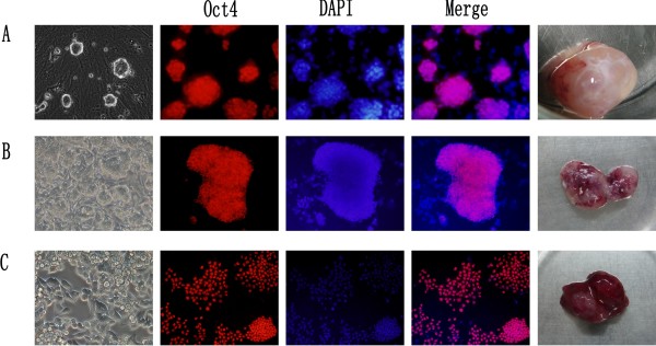
Figure 1.
ESCs, ECCs1 and ECCs2 in 2i medium. (A) ESCs in 2i (left), immunostaining of Oct4 (red), ESCs-converted tumors (right). (B) ECCs1 in 2i (left), immunostaining of Oct4 (red), ECCs1-converted tumors (right). (C) ECCs2 in 2i (left), immunostaining of Oct4 (red), ECCs2-converted tumors (right). DAPI (blue) staining of nuclei for total cell content.

