
Figure 4. UA-induced formation of autophagosomal vaculoes was not followed by autosomal maturation and substrate degradation.
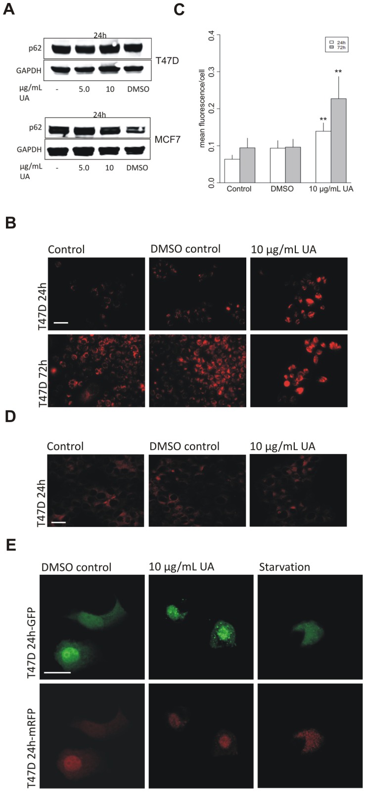
(A) No degradation of p62 was detected, by Western blotting, in T47D and MCF7 cells after treatment with UA (5.0 and 10 µg/mL, 24 h; DMSO 0.1%). (B) Lysotracker, detected by fluorescence microscopy, shows diffuse staining in T47D cells after treatment with UA (10 µg/mL; DMSO 0.2%) for 24 h and 72 h. (C) Lysotracker intensity per cell was quantified by ImageJ and data presented as mean fluorescence value of each group compared to DMSO control. Error bars indicate standard error of the mean, **p<0.001. The scale bar shown represents 100 µm and applies to all panels. (D) No effects on Lamp2 immunostaining were detected, after treatment with UA (10 µg/mL; DMSO 0.2%) for 24 hours. The scale bar shown represents 100 µm and applies to all panels. (E) A plasmid expressing mRFP-GFP-LC3 was transfected into T47D cells. Lack of autophagolysosomal acidification was seen after treatment with UA (10 µg/mL; DMSO 0.2%) for 24 hours by detection of distinct GFP puncta. The scale bar shown represents 20 µm and applies to all panels.