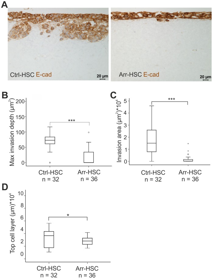
Figure 3. Arresten efficiently inhibits HSC-3 carcinoma cell invasion in an organotypic model.
A. Ctrl-HSC and Arr-HSC cells (7×10⁵) were cultured on top of a collagen gel embedded with human gingival fibroblasts (7×10⁵). The organotypic sections were stained with E-cadherin antibody (brown). Scale bar 20 µm. Tumor cell invasion and growth were quantified by measuring the maximal invasion depth (B), invasion area (C) and area of the top cell layer of pancytokeratin stained sections (D). Mann-Whitney U-test, ***p<0.001, *p<0.05, (n = total number of fields analyzed, 4–5 fields per organotypic section).

