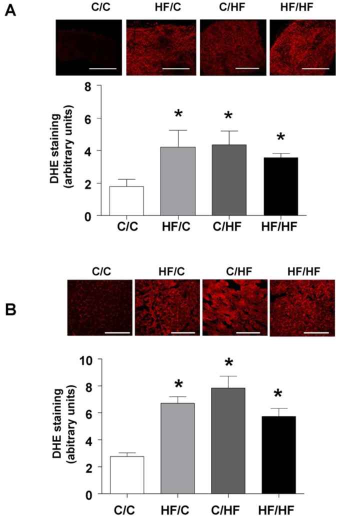
Figure 6. Impact of maternal HF-feeding on offspring redox regulation.
Superoxide generation in (A) fresh femoral artery segments from 15 weeks (n = 4 per group) and (B) vastus muscle from 30 week old offspring (n = 5 per group) assessed using the redox-sensitive dye dihydroethidium (DHE) staining (5 µM in PBS). Representative confocal images obtained from femoral artery and vastus segments from the four offspring groups are shown above each bar graph. Scale bar = 250 µm. Bar graphs represent mean ± SEM. Statistical comparisons were by ANOVA for the effects of maternal and offspring diet followed by comparisons using Dunnett’s Multiple Comparison Test for HF/C, C/HF and HF/HF vs. C/C. * p<0.05).

