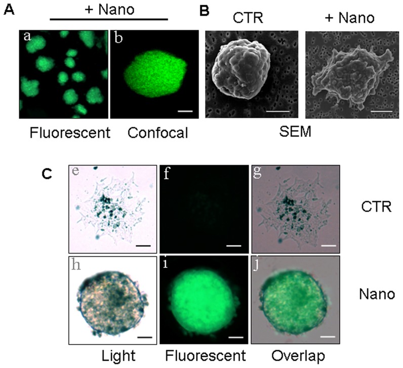
Figure 2. Nanoparticle coating of mouse islets.
(A) Islets incubated with PEG plus coumarin-6 (green)-labeled nanoparticles (Nano) observed under fluorescence (left) and confocal (right) microscopes. Scale bar, 50 µm. (B) Naked control islets (CTR), or pegylated islets coated with coumarin-6 labeled nanoparticles (Nano) imaged by SEM immediately after encapsulation. Scale bar, 100 µm. (C) Islets imaged at 21 days post culture: images e–g show naked islets (CTR), images h–j show islets draped with PEG plus coumarin-6-nano (Nano). The naked islets show degradation in marked contrast to the well-preserved nano-pegylated islets. Scale bar, 50 µm.

