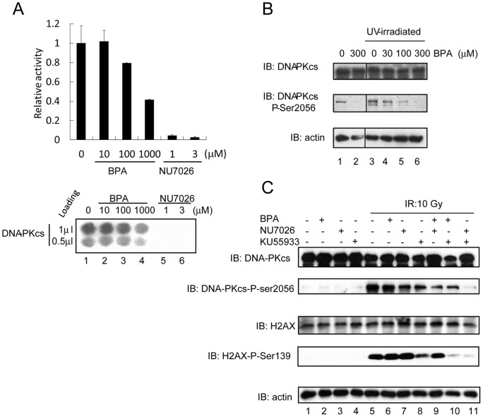
Figure 3. Inhibitory effect of DNA-PKcs kinase activity by BPA.
(A) Protein kinase activity was assayed in vitro using the SignaTECT®. Purified DNA-PKcs, biotinylated p53-derived peptide substrate, DNA, and [γ-32P] ATP were mixed and BPA or NU7026 was added as indicated. Samples were incubated at 30°C for 5 min. Incorporation of 32P into the substrate was measured using an Imaging plate. (B) 293T cells were exposed to 150 J/m2 of 254 nm UV followed by incubation at 37°C for 6 h. Cells were harvested and immunoblot analysis was performed using antibodies to DNA-PKcs-phospho-serine 2056, total DNA-PKcs, and actin (loading control). BPA was added 1 h prior to UV-irradiation at the concentrations indicated. (C) M059K cells were exposed to 10 Gy of γ-rays. Cells were pre-incubated with BPA (300 μM, 3 h), Nu7026 (10 μM, 24 h) or Ku55933 (10 μM, 1 h) separately or in combination. One hour after irradiation, cells were harvested and the amount of DNA-PKcs, DNA-PKcs-phospho-serine 2056, H2AX, H2AX-phospho-serine 139, and actin (loading control) were analyzed by immunoblotting.

