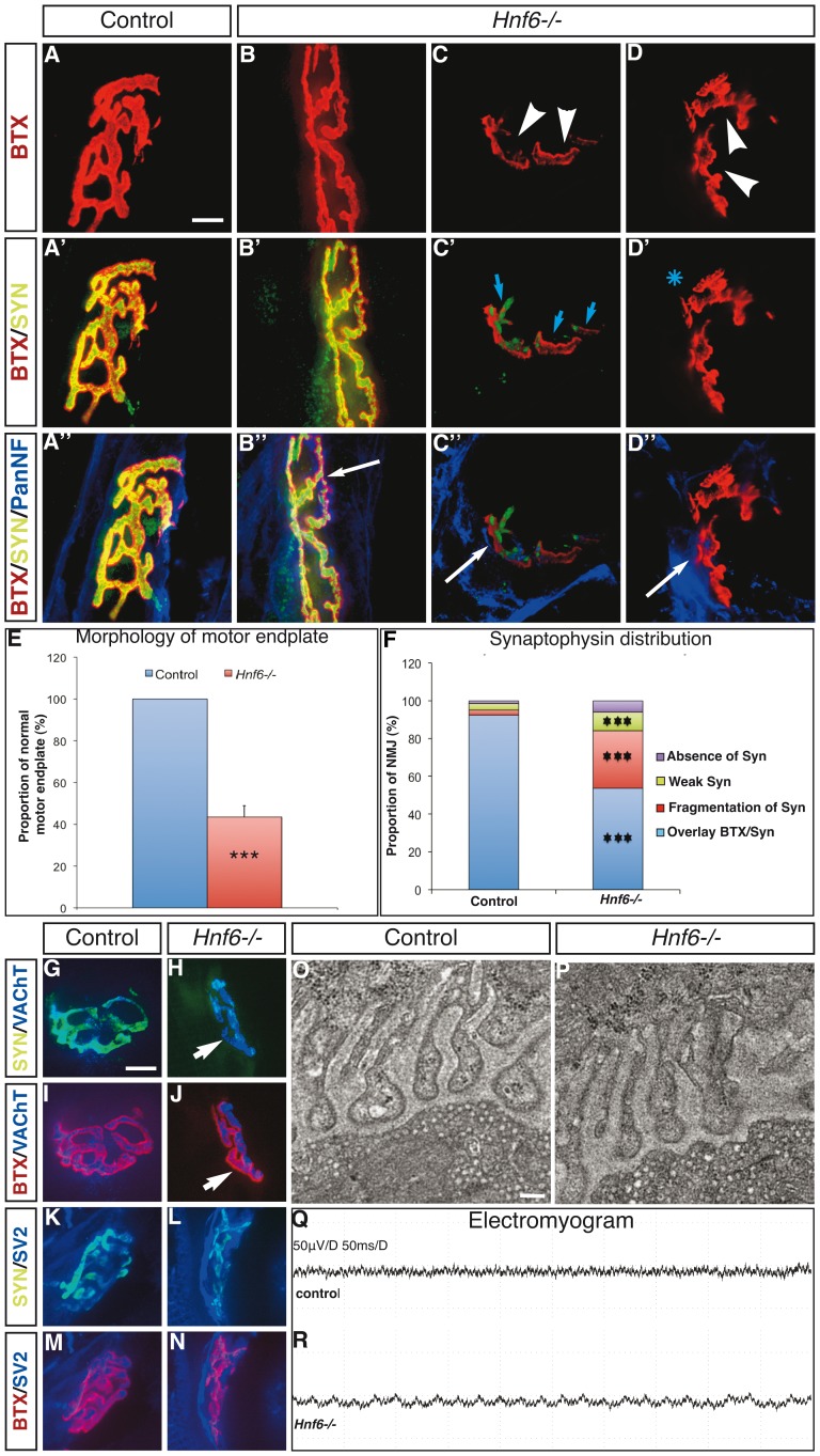
Figure 3. HNF-6 is required for proper morphology of the NMJ.
A–D” , Labeling of acetylcholine receptors by α-bungarotoxin (red) and immunofluorescence detection of synaptophysin (green) and of neurofilaments (blue) on hindlimb sections of control (A–A”) or Hnf6−/− (B–D”) mice. (A–A”) In control mice, NMJ display the expected “pretzel-like” shape, are innervated and show perfect apposition of the synaptophysin labeling to the motor endplate. (B–B”) Some NMJ in Hnf6−/− mice are very similar to that observed in control animals. (C–D”) However, a majority of the Hnf6−/− junctions are characterized by disorganized topology, fragmentation or absence of gutters (arrowheads). These junctions also show defective localization of synaptophysin, which is either absent (asterisk) or fragmented (blue arrows) and ectopically located. In contrast, all the junctions were innervated (white arrows). E , Quantification of motor endplates that exhibit the expected “pretzel-like” unfragmented morphology in control (blue) and in Hnf6−/− (red) mice (n = 6). F, Quantification of defective synaptophysin localization in Hnf6−/− mice. Synaptophysin is either properly superposed to the α-bungarotoxin labeling, as observed in a vast majority of control junctions (blue), or fragmented (red), weak (green) or absent (purple) (n = 5). G–N , Labeling of control (G,I,K,M) or Hnf6−/− (H,J,L,N) junctions with synaptophysin (G,H,K,L, green) or α-bungarotoxin (I,J,M,N, red) and VAChT (G–J, blue) or SV2 (K–N, blue). In Hnf6−/− mice, the VAChT and SV2 are properly localized at the motor terminal ends. O, P , Ultrastructure of the NMJ observed by transmission electron microscopy in control (O) or Hnf6−/− (P) tibialis anterior muscles. Synaptic vesicles are present and normally localized (n = 3). Q, R , Electromyogram recordings in control (Q) or Hnf6−/− (R) mice do not evidence any sign of denervation of the mutant hindlimb muscles (n = 3). BTX: α-bungarotoxin; PanNF: Pan-Neurofilament; SV2: synaptic vesicle 2; SYN: synaptophysine; VAChT: vesicular acetylcholine transporter. Student’s t-test; *** = p<0.001. Scale bar in A and G = 5 µm; scale bar in O = 0.4 µm.

