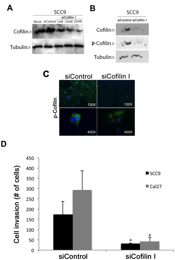
Figure 4. siRNA-mediated knockdown of cofilin-1 resulted in decreased invasive ability of oral cancer cells.
Western blot analysis showed reduced levels of (A) cofilin-1 in SCC-9 cells transfected with different concentrations of siRNA (siCofilin I) for 48 h and of (B) cofilin-1 and p-cofilin in SCC-9 cells transfected with 20 nM siCofilin I for 48 h. (C) Immunofluorescence analysis of cofilin-1 knockdown SCC-9 cells (siCofilin I) using anti-p-cofilin antibody (green). (D) Invasion assays using Matrigel-coated filters were performed on SCC-9 and Cal 27 cells (cofilin-1 knockdown cells and controls). Bar graph represents the mean ± S.E. of the number cells that invaded through the Matrigel from three independent experiments (Student’s t test, * = p<0.01).

