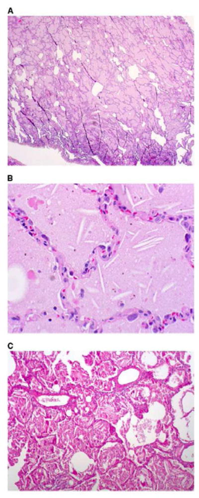
Figure 2.

Histopathological sections of lung biopsy, hematoxylin and eosin stain. (A) Low-power overview showing filling of alveolar spaces by eosinophilic material (magnification 310). (B) High-power view showing granular eosinophilic material and cholesterol clefts. (magnification 3200). Birefrigent particles were identified with polarizing microscopy, consistent with the presence of crystalline indium-tin oxide. (C) Periodic acid-Schiff (PAS) stain after diastase digestion, showing granular, PAS-positive intraalveolar material, and cholesterol clefts (magnification X100). from Cummings, K. Donat W. Ettensohn, David. Pulmonary Alveolar Proteinosis at an Indium Processing Facility. Am J Respir Crit Care Med. 181; 2010: 458–464, with permission).
