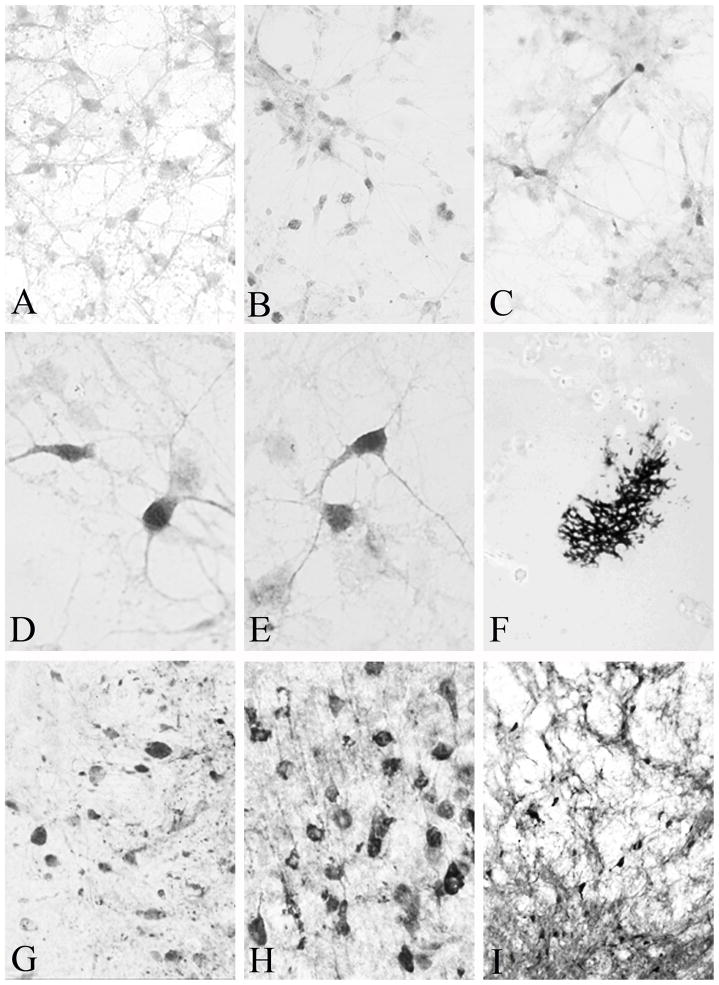
Figure 3.
(A) Exposure of neuronal cultures to the control AAV-NSE-LacZ vector did not result in expression of calbindin immunoreactivity. Only background staining was seen. In contrast, exposure of cultures to AAV-ACP-CB (B) or AAV-NSE-CB (C) resulted in robust expression of calbindin in neurons after 37 hours, visible at low power. (D & E) High power photomicrographs clearly demonstrate dark calbindin immunoreactivity, frequently seen in cultures exposed to AAV-NSE-CB. (F) β-galactosidase staining in the nucleus basalis of Meynert following a 10 μl injection of AAV-NSE-LacZ. This low-power micrograph demonstrates the extent of spread of the injected AAV. High (G and H) and low (I) power micrographs demonstrating calbindin expression in the lateral aspects of the thalamus (G), parietal cortex (H) and globus pallidus/internal capsule component of the nucleus basalis of Meynert 15 days following injection of 5μl of AAV-NSE-CB. Magnification in A-C is 40X, in D & E 60X, in F 4X, in G & H 40X, and in I 10X.

