
Figure 7.
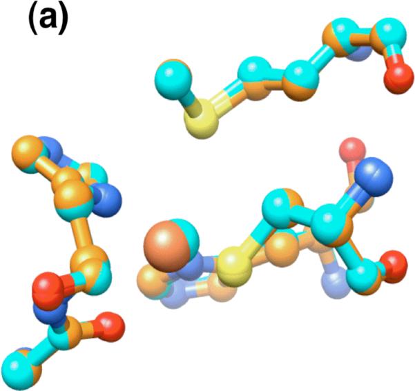

The metal site in the X-ray crystal structure of wild-type iron azurin. (a) Overlay of iron(II) (orange) and copper(II) (blue) azurins. (b) Overlay of chain B of iron(II) azurin (orange) with chain A of apo-azurin (white). The water molecule in apo-azurin is indicated. (c) Overlay of chain B of iron(II) azurin with chain B of apo-azurin.