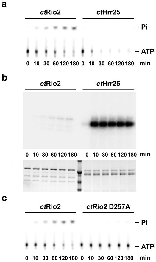
Figure 2. ctRio2 has ATPase activity in vitro.
The amount of released free phosphate (Pi) (a and c) or phosphorylated protein (b) was analyzed in single-turnover experiments using 1μM of the indicated purified recombinant protein. (a) and (c) ATP and Pi were separated by thin-layer chromatography. (b) the amount of phosphorylated protein was analyzed by SDS-PAGE. Dried gels and TLC plates were exposed overnight on a phosphorimager screen (see experimental procedures for a full description). Note that 100 times more of the reactions were loaded in panel b in comparison to panel a and c.

