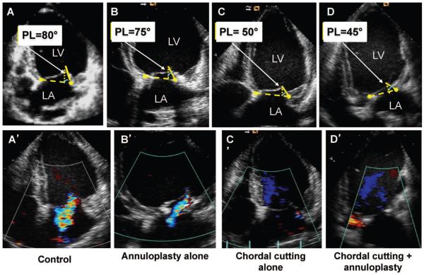
Figure 2.
Midsystolic apical 2-dimensional echocardiographic images. Mitral valve measurements were performed at midsystole. A, Leaflet apical tenting relative to the annulus with a prominent bend in the basal anterior leaflet and markedly limited posterior leaflet (PL) motion, with PL angle relative to annulus of 80°. A’, Control with moderate mitral regurgitation (MR) with a central jet into the left atrium. B, Ring alone with concave anterior leaflet (toward the left ventricle [LV]) with restricted motion of the PL (due to the ring) and PL angle of 75°. B’, Ring alone with mild MR. C, Chordal cutting alone with less LV remodeling and a decrease of PL angle to 50° and concave anterior leaflet. C’, Minimal MR. D. Chordal cutting plus ring. Less LV remodeling with a decrease of PL angle to 45° and more concave anterior leaflet is shown. D’, No MR (bottom). LA indicates left atrium.

