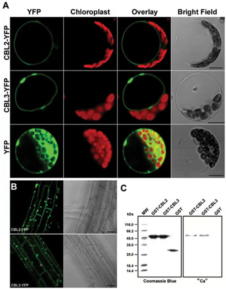
Figure 2.
Subcellular localization and Ca2+ binding of CBL2 and CBL3. (A) Confocal microscopy analysis of YFP signals from Arabidopsis mesophyll protoplasts transiently expressing CBL2-YFP and CBL3-YFP fusions or YFP alone as indicated. In each panel, the YFP signals (green), chloroplast fluorescence (red), overlay (green plus red) and bright field images from the same cell are shown. Scale bar = 10 μm. (B) CBL2/3-associated YFP signals in mature root cells of transgenic Arabidopsis plants stably expressing CBL2-YFP (top panel) or CBL3-YFP (bottom panel). Arrows point to vacuolar membrane invagination away from the plasma membrane. (C) Purified GST-CBL2 and GST-CBL3 fusion proteins as well as GST control were separated on 12.5% SDS-polyacrylamide gel by electrophoresis and stained with Coomassie Blue (left panel) or electroblotted onto a nitrocellulose membrane, incubated with 45Ca2+ and autoradiographed (right panel).

