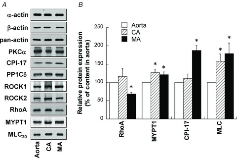
Figure 12. Expression of various contractile and regulatory proteins in rat small mesenteric artery (MA), caudal artery (CA) and aorta.

Protein contents in extracts were adjusted by equalizing smooth muscle-specific α-actin content between different arteries. A, representative Western blot images of various proteins in rat aorta, CA and MA. B, average expression levels of proteins, the contents of which were significantly different between arteries (P < 0.05). n= 6–30.
