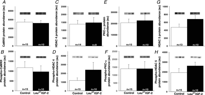
Figure 4. The increase in the area of binucleated cardiomyocytes in the left ventricle was not stimulated through activation of the Gαq signalling pathway.

There were no differences in the amounts of CaMKII (A), phospho-CaMKII (B), HDAC 4 (C), phospho-HDAC 4 (D), PKC-α (E), phospho-PKC-α (F), HDAC 5 (G) and phospho-HDAC 5 (H) proteins in control compared with Leu27IGF-2-infused fetuses. Representative Western blots from 3 animals in each group for each protein. Sample size for each group is indicated in the bar.
