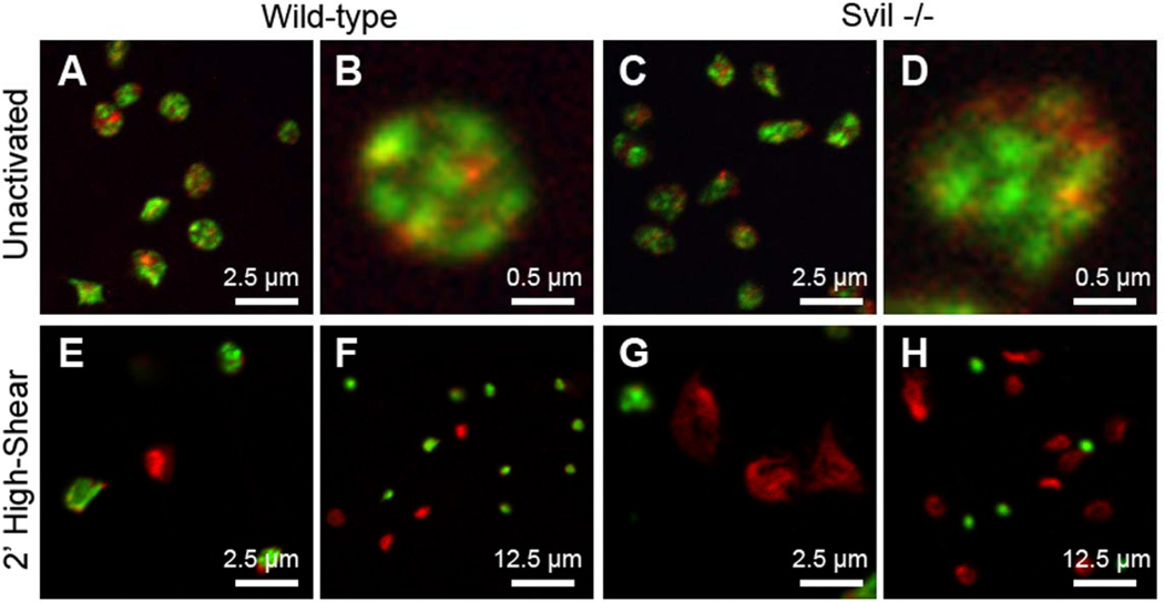
Figure 7.
Immunofluorescence micrographs of (A, B, E, F) wild-type or (C, D, G, H) supervillin-deficient platelets fixed (A–D) statically onto glass or (E–H) after activation for 2 min on collagen under high-shear (1200 s−1) flow. F-actin (red) was visualized with AlexaFluor568-phalloidin; myosin IIA (green) was stained with antibody. Bars, 2.5 µm, 0.5 µm, and 12.5 µm, as indicated.

