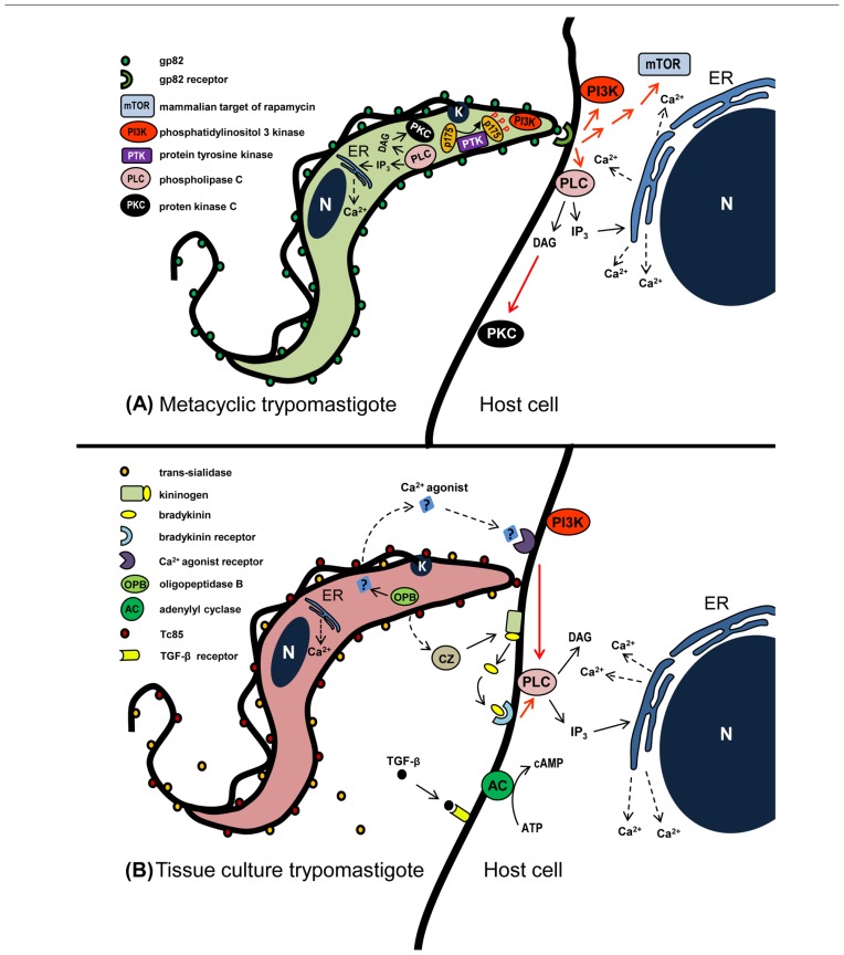
FIGURE 1.
Schematic representation of signaling molecules and pathways that may be activated during T. cruzi entry into non-phagocytic mammalian cells. (A) In metacyclic forms that enter host cells in gp82-mediated manner, activation of PLC generates DAG and IP3. DAG stimulates PKC and IP3 promotes Ca2+ release from IP3-sensitive compartments. PI3K and PTK are also activated, the latter mediates phosphorylation of p175. In the host cell, the recognition of gp82 by its receptor triggers the activation of PI3K, mTOR, and PLC, the latter generating DAG and IP3. DAG stimulates PKC and IP3 promotes Ca2+ release from endoplasmic reticulum (ER). (B) During TCT interaction with host cells, a Ca2+ agonist generated by parasite OPB binds to its receptor and triggers PLC activation. Then IP3-mediated Ca2+ release from ER ensues. Bradykinin, produced from kininogen by the action of TCT cruzipain, binds to bradykinin receptor and triggers PLC activation. Red arrows indicate activation, possibly not directly, but through as one or more as yet undefined elements.

