
Figure 1.
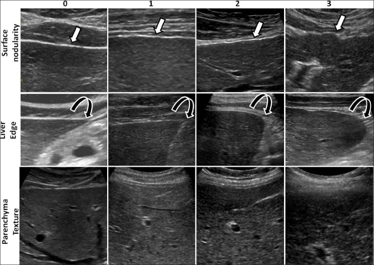
Examples of ultrasound images from different patients illustrating the ultrasound features evaluated and scores assigned to the features. In the top row are images showing the liver surface demonstrating smooth to severe nodularity (arrows) and in the middle row, images showing the liver edges from sharp to rounded edge (curved arrows). In the bottom row, note the change in fine granular echoes in normal liver to severely coarse echoes in cirrhotic liver.