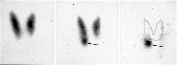
Figure 5.
Example of parathyroid scintigraphy according to the dual-tracer protocol (99m-Tc-pertechnetate and 99mTc-sestamibi). (On the left) the 99 mTc-pertechnetate scan shows a normal thyroid gland. (In the center) the 99mTc-sestamibi scan suggests the presence of adenoma of the lower right parathyroid glaand (arrow), better outlined when subtracting the 99mTc-pertechnetate scan from the summation scan (arrow).

