Figure 3.

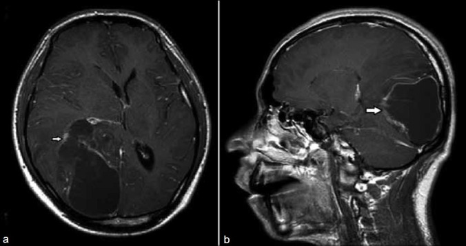
(a) Gd contrast enhanced T1 weighted axial image at the level of caudate nucleus shows the lesion wall enhancement (arrow). (b) Corresponding sagittal plane lesion involving temporo-occipital lobe reconfirms the axial plane features

(a) Gd contrast enhanced T1 weighted axial image at the level of caudate nucleus shows the lesion wall enhancement (arrow). (b) Corresponding sagittal plane lesion involving temporo-occipital lobe reconfirms the axial plane features