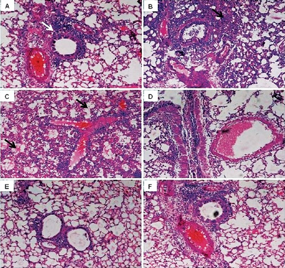
Fig. 4.

Lung histopathology of mice after infection with influenza virus. Lungs were fixed, sectioned, and stained with haematoxylin and eosin. A-C: Tissue from mice infected with influenza virus alone showing increase in infiltration of inflammatory cells into alveolar walls (white arrow head in A) with focal areas of consolidation (black arrow head in B) and necrosis of epithelial cells (arrows in C) on day 3, 5 and 7 p.i.; D-F: Tissue from mice simultaneously administered with rTGF-β1 showing less involvement of bronchioles and infiltration of inflammatory cells and normal parenchyma on day 3, 5 and 7.
