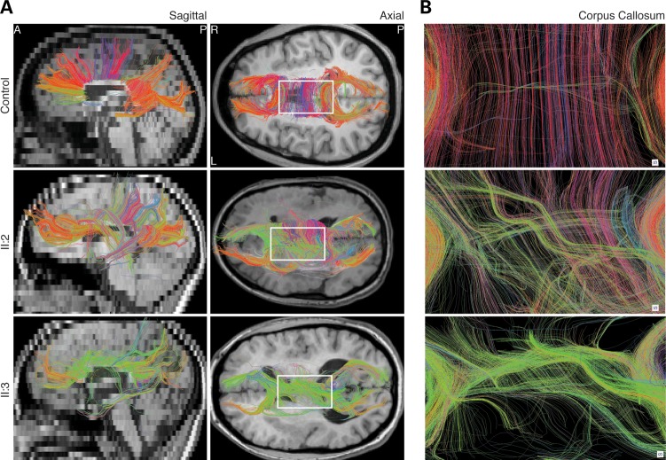
Figure 3.
Abnormal homotopic connectivity associated with TUBB2B-E421K. Human DTI images segmenting commissural fibers of the CC. (A) Sagittal and axial views show that TUBB2B-E421K patients have a paucity of commissural fibers in the CC body, consistent with structural MRI findings. Furthermore, while many fibers in the healthy control are reconstructed with red color, many commissural fibers in TUBB2B-E421K patients are colored green, indicating a lack of normal homotopic connectivity. (B) Zoom in of the boxed region in (A) confirms that homotopic connectivity is disrupted in patients. CC, corpus callosum. Color coding: red, left–right; green, anterior–posterior; blue, superior–inferior.

