
Figure 1. LSB3i treated hPSCs rapidly acquire a nociceptor phenotype within 12 days.
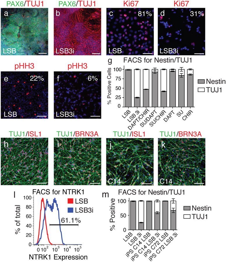
Upon staining for TUJ1, a neuron marker, (a) compared to LSB alone, (b) far greater numbers of positive cells are observed when three inhibitors (CHIR99021, DAPT, SU5402; termed 3i) are applied 48 hours after treatment of hPSCs with LSB. Comparing expression of Ki67 and phospho-histone H3 in LSB (c,e) and LSB3i (d,f) treated hPSCs indicated a stark decline in proliferation by day 12. (g) Intracellular antibody FACS staining for Nestin and TUJ1 indicated a stark contrast in the number of neurons generated by LSB (2% TUJ1+) compared to LSB3i (75% TUJ1+). When one or two of the three inhibitors used in 3i are added, the same level of TUJ1 cells is not achieved, however CHIR with either SU5402 or DAPT can achieve greater than 53% neurons, indicating a requirement for CHIR in the formation of TUJ1+ neurons. TUJ1 positive neurons from LSB3i treated hPSCs express (h) ISL1, (i) BRN3A. (j,k) Similar neurons are observed when hiPSC lines are treated with LSB3i (C14 shown). (l) Greater than 61% of all cells express NTRK1 measured by FACS. (m) LSB3i treated hiPSCs form neurons at a moderate efficiency. Scale bars for (a,b) are 200 μm and (c-f,h-k) are 100 μm.