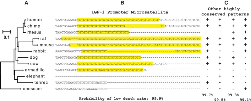
Fig. 1.—
Conservation of a promoter microsatellite. (A) Phylogeny of mammalian species used in our analyses. Branch lengths are measured in average number of substitutions per 4-fold degenerate site. (B) An example of a highly conserved microsatellite (highlighted in yellow) uncovered by the birth–death mixture model. This GT-motif microsatellite is found ∼800 bp upstream of the insulin growth factor 1 (igf1) transcription start site. (C) Generic examples of other patterns of highly conserved loci. The + and − symbols represent present and absent, respectively.

