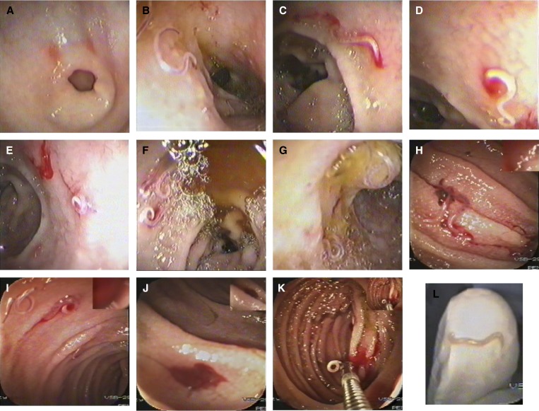
Figure 1.
Serial endoscopic imaging of ancylostomiasis-induced gastrointestinal bleeding. (A) Gastric erosions around the pyloric ring. (B) Worms in the duodenal bulb with nearby red spots of previous bites. (C) A duodenal bulb erosion at the worm's attachment site. (D) A close-up view on a bleeding spot at the attachment site. (E) A coiled worm at the end of the duodenal first part with a nearby fresh blood. (F) Several worms in the duodenal second part with bile around. (G) A worm coiled over the ampulla of Vater. (H) Worms in the jejunum with surrounding clotted blood. (I) Jejunal worms with surrounding fresh blood. (J) A blood pool in a jejunal segment of previous bites. (K) A worm caught by the biopsy forceps with dark and fresh blood spots in the jejunum. (L) A close-up view of the extracted worm on the endoscopist's gloved index finger tip (imaged by the videoendoscope itself).

