Fig. 1.
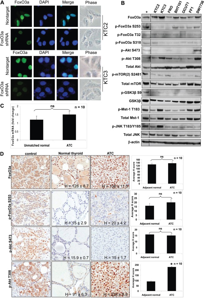
FoxO3a is predominantly nuclear in ATC cell lines. (A) ICC shows nuclear localization of FoxO3a in KTC2 and KTC3 cells. Cells were lentiviral-infected with either nontarget or FoxO3a 1566 shRNA. Less fluorescence is seen in the FoxO3a-silenced cells, indicating that the nuclear expression is specific. DAPI was used to stain the nuclei. (B) Western blot of ATC cell lines for total and phosphorylated forms of FoxO3a, Akt, mTOR, Mst-1 and JNK expression. MDA231 was used as a positive control (+) that was run on the same blot (another control was removed). β-actin was used as a loading control. (C) QPCR of unmatched normal and ATC patient tissues shows that there is no significant difference between FoxO3a mRNA levels. Data are plotted as average fold change compared with unmatched normal ± s.d. (n = 10). (D) IHC in adjacent normal thyroid and ATC patient tissues demonstrates FoxO3a is nuclear and expression is similar for both as shown by H scores. Normal patient pancreatic tissue was used as a positive control, and shows cytoplasmic staining of FoxO3a, further demonstrating the specificity of the antibody for nuclear staining in thyroid tissue (top left panel). Breast tumor tissue was used as a positive control for the remaining control panels.

