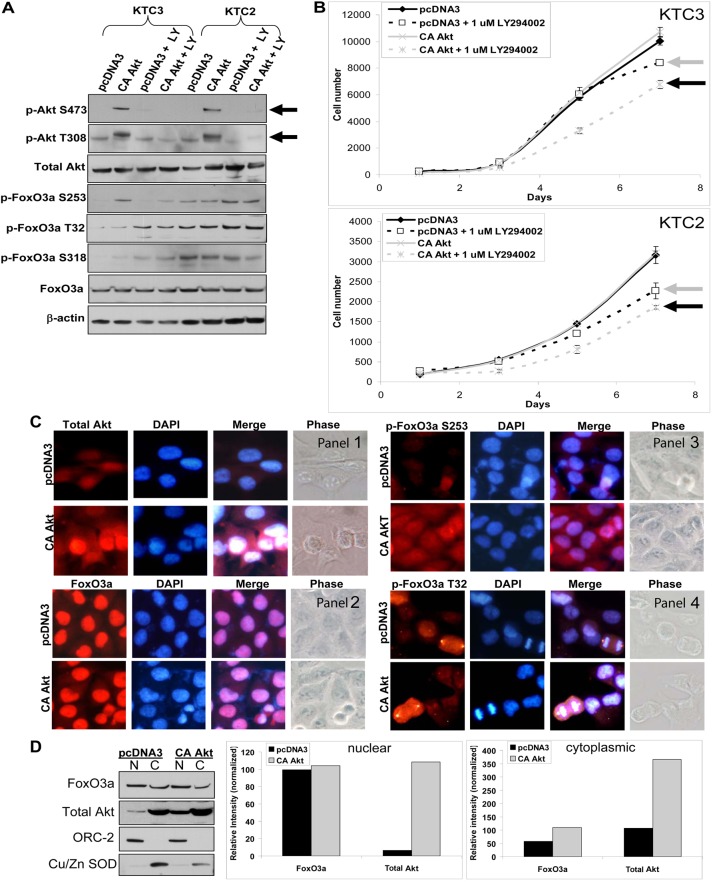
Fig. 2.
Overexpression of constitutively active Akt has no effect on proliferation. (A) Western blot of KTC3 and KTC2 cells transfected with pcDNA3 control or a constitutively active (CA) Akt expression plasmid demonstrates overexpression of CA Akt versus pcDNA3 control. When cells were untreated or treated with 1 µM LY294002 (LY), it was shown that LY attenuates p-Akt levels. Total FoxO3a and p-FoxO3a protein levels were also examined for each of these conditions. (B) Proliferation curves of transfected KTC3 and KTC2 cells over 7 days, show that there is no difference in proliferation rates between the pcDNA3 control and the CA Akt cells. Treatment with the PI3-K inhibitor, LY294002, resulted in only slight growth inhibition in the pcDNA3 controls (gray arrow) compared with pcDNA3-DMSO-treated cells, whereas the CA Akt cells showed modest but statistically significant growth inhibition (black arrow) when compared with the LY-treated pcDNA3 cells of both cell lines. Data are plotted as cell number ± s.d. (C) ICC of transfected KTC3 reveals that total Akt is both nuclear and cytoplasmic (panel 1). Total FoxO3a (panel 2) remains predominantly nuclear in the CA Akt cells with minimal shuttling of p-FoxO3a S253 to the cytoplasm (panel 3), whereas p-FoxO3a T32 remains cytoplasmic (panel 4). DAPI was used to stain the nuclei. (D) Subcellular fractionation followed by western blotting and densitometric analysis confirmed that FoxO3a is predominantly in the nuclear fraction and when CA Akt is overexpressed, very little if any FoxO3a is shuttled into the cytoplasm. ORC-2 was used to normalize for the nuclear fraction and Cu/Zn SOD for the cytoplasmic fraction. N, nuclear, C, cytoplasmic.

