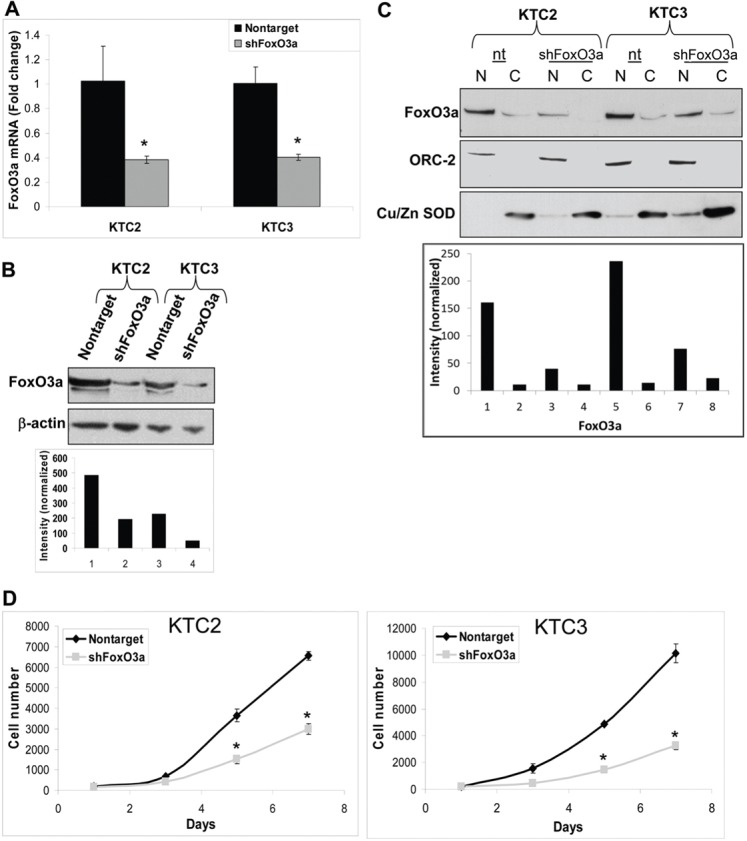
Fig. 3.

Silencing FoxO3a reduces proliferation. (A) QPCR verifies that FoxO3a has been silenced in KTC2 and KTC3 cells that have been infected with lentiviral FoxO3a 1566 shRNA but not in nontarget control cells. Data are plotted as fold change ± s.d. as compared with nontarget control; *P<0.05. (B) Western blot of the same cells confirms that FoxO3a is reduced in the FoxO3a shRNA cells compared with nontarget control. β-actin was used as a loading control and was used to normalize data for densitometric analysis. (C) The same cells were then fractionated to demonstrate that FoxO3a is predominantly in the nuclear fraction and its expression can be attenuated by shRNA. ORC-2 and Cu/Zn SOD were used to normalize data for densitometric analysis. N, nuclear; C, cytoplasmic. (D) Proliferation curve of the lentiviral-infected cells showing decreased proliferation over 7 days when FoxO3a is silenced. Data are plotted as cell number ± s.d. and *P<0.05, compared with nontarget control.
