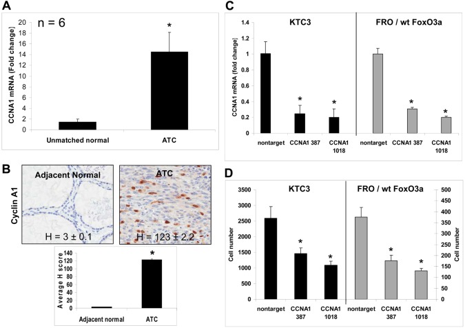
Fig. 7.
Cyclin A1 is overexpressed in ATC patient tumor tissues. (A) QPCR of unmatched normal and ATC patient tissues show there is a significant increase in CCNA1 mRNA levels. Data are plotted as average fold change compared with unmatched normal ± s.d., *P<0.05. (B) IHC in adjacent normal and ATC patient tissues demonstrates cyclin A1 is overexpressed as shown by H scores. (C) QPCR verifies that CCNA1 mRNA has been silenced in KTC3 and FRO/wt FoxO3a cells that have been infected with lentiviral CCNA1 387 and 1018 shRNA compared with levels in nontarget control. Data are plotted as fold change ± s.d. compared with nontarget control, *P<0.05 (D) Proliferation analysis of lentiviral-infected cells shows that when CCNA1 is silenced in KTC3 or FRO/wt FoxO3a-overexpressing cells, decreased cell proliferation occurs. Data are plotted as cell number ± s.d.; *P<0.05, compared with nontarget control.

