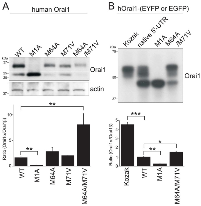
Fig. 2.

Two isoforms of Orai1 are translated by alternative translation initiation. (A) Western blot showing heterogeneously expressed Orai1 proteins in HEK293 cells transfected with the cDNAs of WT, first methionine (M1A) mutant, second methionine (M64A) mutant, third methionine (M71V) mutant and double mutant of second and third methionine (M64A/M71V) mutant of Orai1. (B) Western blot showing the expression of Orai1 proteins tagged with fluorescent protein in HEK293 cells transfected with the cDNAs of WT with Kozak sequence, WT with native 5′-UTR, M1A mutant and M64A/M71V double mutant of Orai1. Cell lysates were treated with PNGaseF overnight and resolved by 4–20% gradient (A) or 10% (B) SDS-PAGE. The blots were probed with anti-Orai1 antibody and anti-actin antibody for loading control. Representative data from at least three independent experiments are shown. Lower panels represent the ratio of Orai1α and Orai1β calculated by densitometric analysis. Data are means ± s.e.m.; *P<0.05, **P<0.01, ***P<0.001, one-way ANOVA followed by Tukey's test (n = 3).
