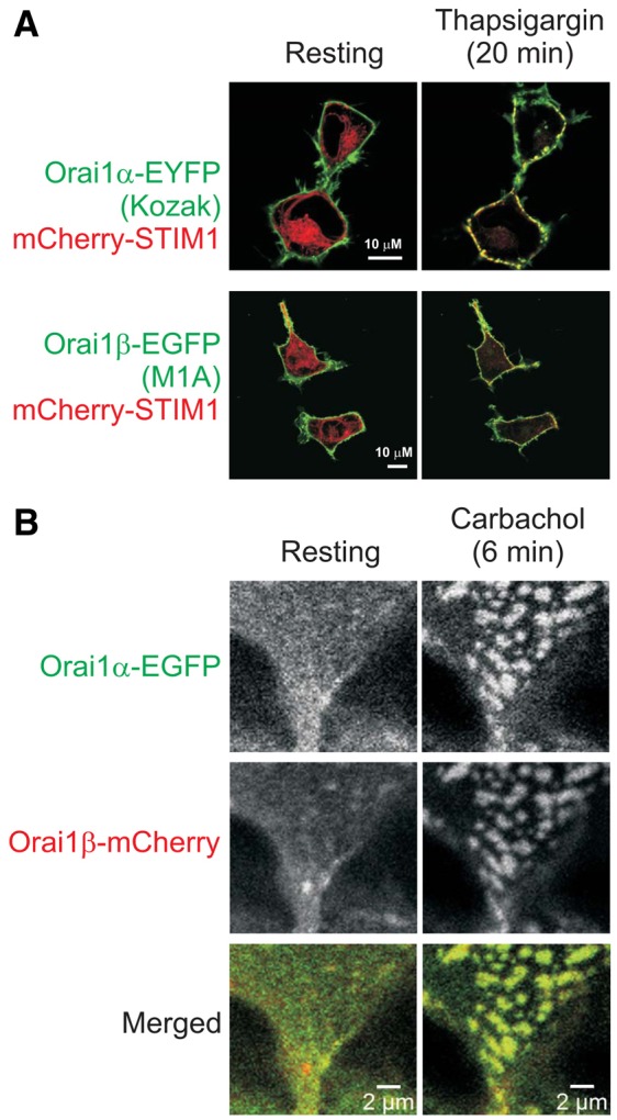
Fig. 3.

Both isoforms of Orai1 protein localize predominantly to the plasma membrane and translocate to puncta upon store-depletion. (A) Confocal images of HEK293 cells co-expressing Orai1-EYFP or Orai1-EGFP with mutations that result in expression of exclusively Orai1α or Orai1β forms, together with mCherry-STIM1. Confocal images were taken every 5 minutes. After collecting the first image, cells were treated with thapsigargin in the absence of extracellular Ca2+. The images taken just prior to, and 20 minutes after thapsigargin treatment are shown and are representative of at least three independent experiments. (B) Confocal images of HEK293 cells co-expressing Orai1α-EGFP and Orai1β-mCherry before and 6 minutes after store depletion by 0.5 mM carbachol. Results are representative of four independent experiments.
