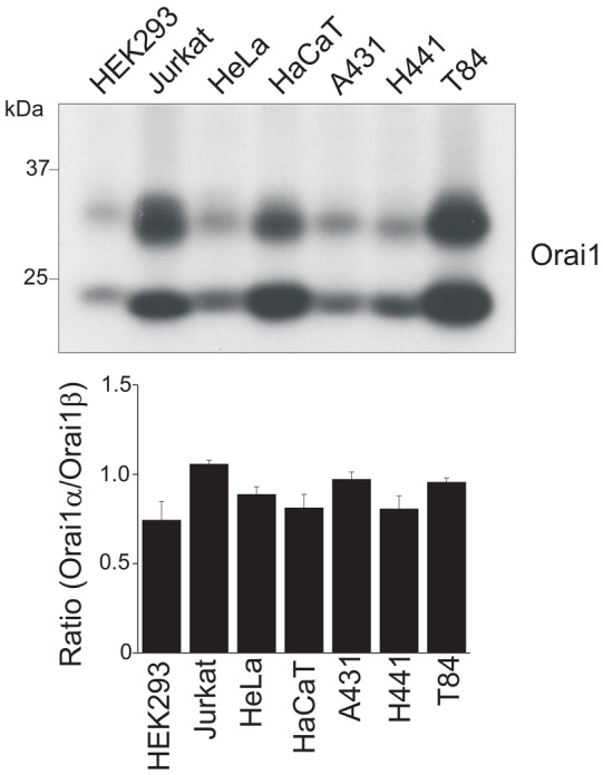
Fig. 6.

Expression of two isoforms of Orai1 in various human cell lines. Western blot showing the expression of endogenous Orai1 in HEK293 cells, Jurkat T cells, HeLa cells, the keratinocyte cell line HaCaT, squamous carcinoma A431 cells, lung adenocarcinoma H441 cells and colorectal adenocarcinoma T84 cells. Cell lysates from each cell type were treated with PNGaseF overnight and dissolved in 10% SDS-PAGE. 15 µg of total protein were loaded into each lane except HaCaT and T84. 7.5 µg of total proteins were loaded into the lanes of HaCaT and T84 due to higher expression of endogenous Orai1 protein these cell lines. The blot was probed with anti-Orai1 antibody. Representative data from three independent experiments are shown. Lower panel represents the ratio of Orai1α and Orai1β calculated by densitometric analysis. Data are means ± s.e.m.
