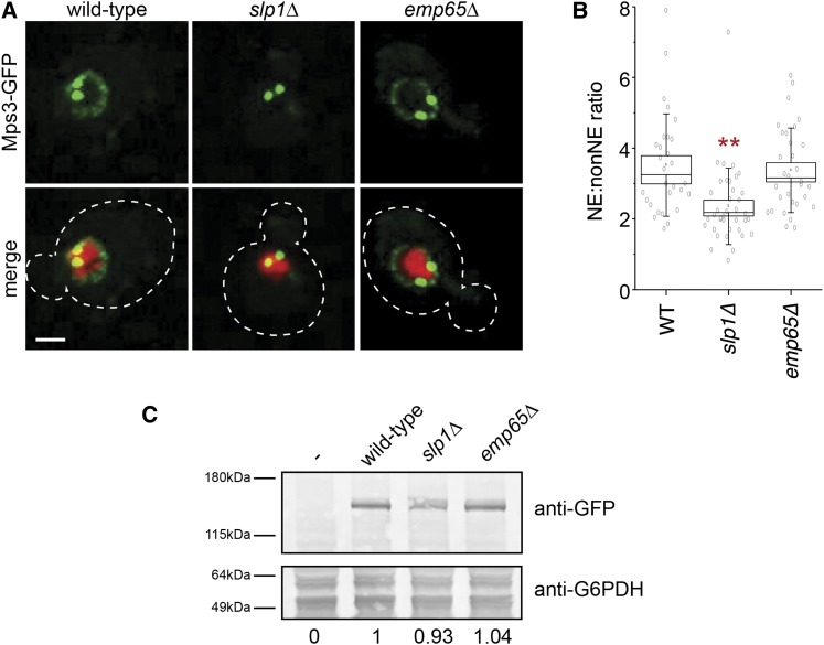
Figure 8 .
Slp1 and Emp65 are required for Mps3 localization to the NE. (A) Localization of Mps3-GFP (green) and H2B-mCherry (red) in wild-type (SLJ5669), slp1Δ (SLJ6783), or emp65Δ (SLJ6785). The cell is outlined in white based on the DIC image. Bar, 2 µm. (B) Quantitation of NE/non-NE ratio in Mps3-GFP in images from (A) was performed as previously described (Gardner et al. 2011). Values for each data point (gray circles), the mean (black square) and median values (line), SE (box) and SD (lines) for each sample are depicted. Values that are statistically significant from wild-type (P < 10−4) are indicated with an asterisk. (C) Protein levels in whole-cell extracts of cells from (A) were determined by western blotting with anti-GFP antibodies. Glucose-6-phosphate dehydrogenase (G6PDH) serves as a loading control and allows for normalization of the levels of Mps3-GFP in different strains (below). The strain lacking Mps3-GFP was assigned a value of 0 whereas the wild-type strain containing Mps3-GFP was given a value of 1.

