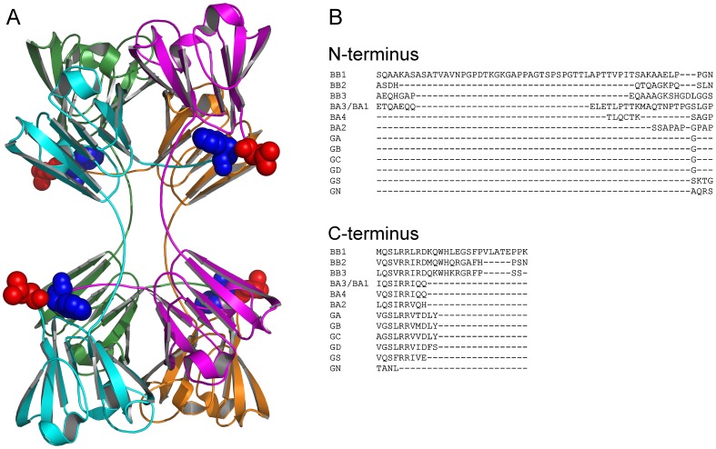
Figure 1. Crystal structure of βB2-crystallin and sequence alignment of β/γ-crystallins.
(A) Crystal structure of βB2-crystallin (PDB ID: 2BB2). The four subunits are labeled in cyan, green, orange and magenta, respectively. Leu15 at the N-terminus (red) and Trp195 at C-terminus (blue) are highlighted by the space-filling model to show the role of N-terminus in tetramerization of βB2-crystallin. (B) Sequence alignment of the N- and C-termini of β/γ-crystallins. The sequence alignment was performed using the online software MAFFT (http://www.ebi.ac.uk/Tools/msa/mafft/). The sequences used for alignment are: βB1-crystallin (BB1, P53674), βB2-crystallin (BB2, P43320), βB3-crystallin (BB3, P26998), βA3/A1-crystallin (BA3/BA1, P05813), βA2-crystallin (BA2, P53672), βA4-crystallin (BA4, P53673), γA-crystallin (GA, P11844), γB-crystallin (GB, P07316), γC-crystallin (GC, P07315), γD-crystallin (GD, P07320), γN-crystallin (GN, Q8WXF5) and γS-crystallin (GS, P22914).

