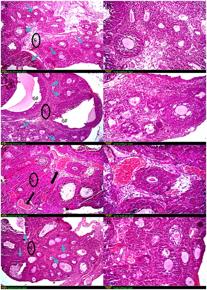
Figure 4. Representative photomicrographs of hematoxylin and eosin-stained ovarian tissue sections.
Histological sections of control (A and B) and sodium selenite (C and D) treated ovaries exhibit similar organization, with different stages of growing follicles (blue arrows). Compared with controls, γ-irradiated ovaries (E and F) have few, if any, resting oocytes in the cortex with severe hemorrhage (black arrow). Many oocytes in small primary follicles are degenerating in irradiated ovaries. (G and H) Sections taken from ovaries of rats exposed to γ-irradiation and pre-treated with sodium selenite show the apparent normal structure of the ovary, with multiple types of ovarian follicles. Scale bar, 10 µm. GF: Graffian follicle, S: Stroma.

