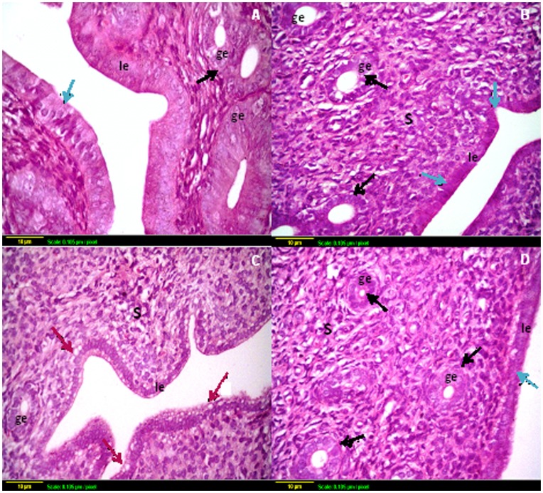
Figure 5. Representative photomicrographs of hematoxylin and eosin-stained uterine tissue sections.
(A) Section taken from uterus of control rat and (B) Section taken from the uterus of sodium selenite treated rat show normal mucosal lining epithelium (blue arrow) with multiple glands (black arrow). (C) Section taken from the uterus of rats subjected to γ-radiation shows high degeneration of the mucosal epithelium with vacuole appearance (red arrow). (D) Section taken from the uterus of rats subjected to γ-radiation and pre-treated with sodium selenite shows regeneration of the glandular and luminal epithelium with intact structure. Scale bar, 10 µm. le: luminal epithelium, ge: glandular epithelium, S: stroma.

