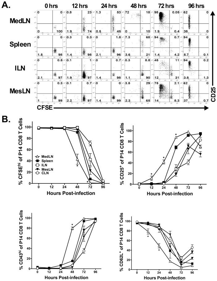
Figure 3. Intraperitoneal infection with LCMV results in rapid antigen display in the MedLN.
CFSE-labeled Thy1.1+ P14 TCR transgenic CD8 T cells were adoptively transferred into naïve Thy1.2+ C57BL/6 mice that were subsequently infected i.p. with LCMV 24 h later. At the indicated times, various organs were harvested and P14 CD8 T cells were assessed for CFSE dilution and expression of activation markers. (A) Representative dot plots showing CFSE dilution profile and CD25 expression. (B) Quantification of CFSE dilution as well as CD25, CD43glyco and CD62L expression. Representative data from one of three individual experiments with three mice at each time point is shown. CD62L data is from two individual experiments with three mice at each time point. *, MedLN is significantly different (p<0.05) as compared to all other tissues as determined by ANOVA with a Tukey post-test. Error bars represent the standard error of the mean for three mice per group.

