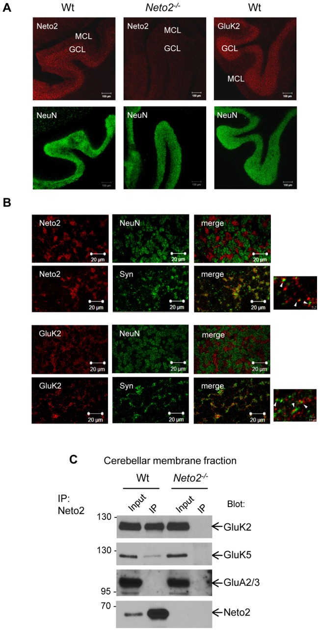
Figure 1. Neto2 is associated with KARs in the cerebellum.

(A) Confocal micrographs of immunostained cerebellar slices. Antibodies used for immunostaining are indicated in the top left corner of each image. In the cerebellum, the NeuN antibody stains the neuronal nuclei of granule cells but does not recognize Purkinje cells. MCL, molecular cell layer; GCL, granule cell layer; Wt, wild-type sections; Neto2−/−, Neto2-null sections. Scale bar, 100 μm. (B) High-magnification confocal microscopy of the cerebellar granule cell layer immunostained with Neto2, GluK2, NeuN, or synaptophysin antibodies. Scale bar, 20 μm; scale bar (small panels on the right), 5 μm (C) Immunoblot of immunoprecipitates from the cerebellum. Blot: antibody used for immunoblot analysis; IP: immunoprecipitate. The input represents 2% of the material used in the immunoprecipitation experiment.
