
Figure 1.
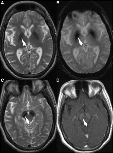
MRI Features Associated with WDR45 Mutations
T2-weighted turbo-spin-echo (TSE) MRI shows (A) a hypointense signal (arrow) in the globus pallidus, which “blooms” on fluid-attenuated inversion-recovery sequence (B, arrow), consistent with iron. A hypointense signal (C, arrow) in the substantia nigra on T2-weighted TSE shows iron, and T1-weighted imaging demonstrates a hyperintense halo (D, arrow) in the substantia nigra.