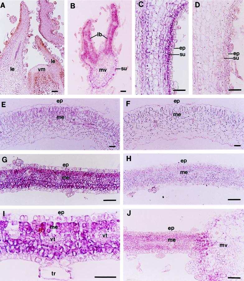
Figure 6.
In situ localization of PNZIP mRNA in different P. nil organs and following various light treatments. Seedlings were grown in continuous light for 6 d and then were exposed to 12 h of darkness or were interrupted at the 8th h by a 10-min NB with red light. Afterward, longitudinal and transverse sections (7 μm thick) through the shoot meristem, hypocotyl, cotyledon, and leaf tissues were collected and hybridized with a PNZIP antisense RNA probe (A–C, E, G, I, and J) or a PNZIP sense RNA probe as a control (D, F, and H). Probes were labeled with digoxigenin-UTP. The transcript-specific hybridization signal is shown in purple. A, Vegetative shoot meristem of a 7-d-old seedling. B, Young expanding leaf taken from the fourth or fifth position below the shoot meristem. C, Hypocotyl. D, Hypocotyl hybridized with a PNZIP sense RNA probe as a control. E, Cotyledon. F, Cotyledon hybridized with a PNZIP sense RNA probe as a control. G, Leaf. H, Leaf hybridized with a PNZIP sense RNA probe as a control. I, Higher magnification of a young leaf. J, Young leaf after an NB treatment. ep, Epidermis; lb, leaf blade; le, leaf; me, mesophyll; mv, main vein; su, subepidermis; tr, trichome; vm, vegetative meristem; and vt, vascular tissue. Bar = 25 μm.

