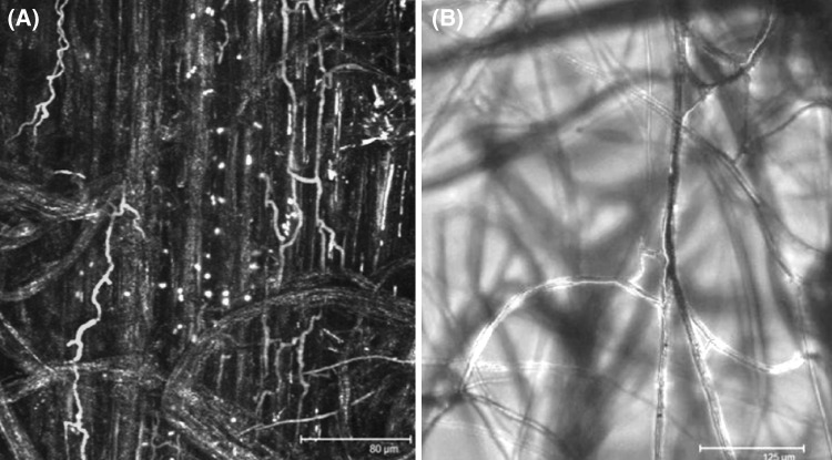
Fig. 1.
Green fluorescent labeled Trichoderma velutinum G1/8 on sterile grown 2 weeks old sugar beet seedlings. Confocal laser scanning microscopy (CLSM) was performed with a Leica TCS SPE confocal microscope (Leica Microsystems, Mannheim, Germany). a Root surface (yellow) and T. velutinum hyphae (green). Hyphae grow between the root cells and follow their cell shape and direction. b Differential interference contrast microscopy of the lateral roots and root hairs combined with CLSM of green T. velutinum hyphae following the growth direction of the root hairs. (Color figure online)

