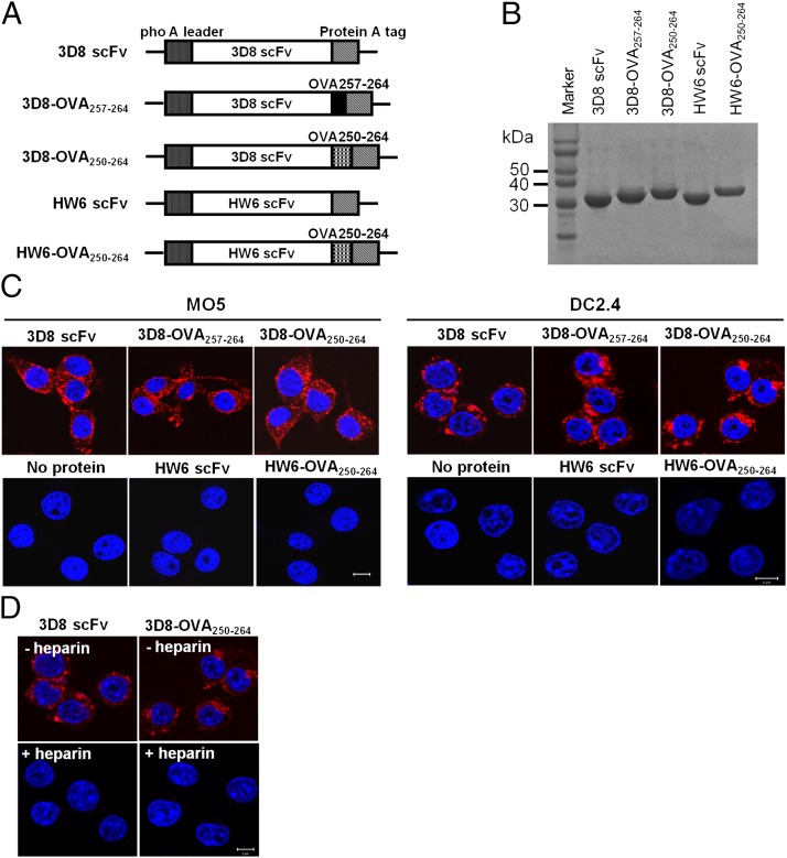
FIGURE 1.
Production of 3D8-OVA fusion proteins and their internalization by MO5 and DC2.4 cells. (A) Schematic diagram showing the expression vectors for the indicated proteins. (B) SDS-PAGE of purified proteins. Proteins (∼20 μg) were separated on 12% SDS-PAGE gels and visualized with Coomassie blue. (C and D) Cell-penetrating activity of proteins. MO5 and DC2.4 cells were incubated with the indicated proteins (10 μM) for 6 h at 37°C in the absence (C) or presence (D) of heparin (50 IU/ml). After washing, fixation, and permeabilization, the cells were incubated with rabbit IgG, followed by TRITC-labeled anti-rabbit IgG Ab (red). Nuclei were stained with Hoechst 33342 prior to analyses under a confocal microscope. Scale bar, 10 μm. Data are representative of three independent experiments.

