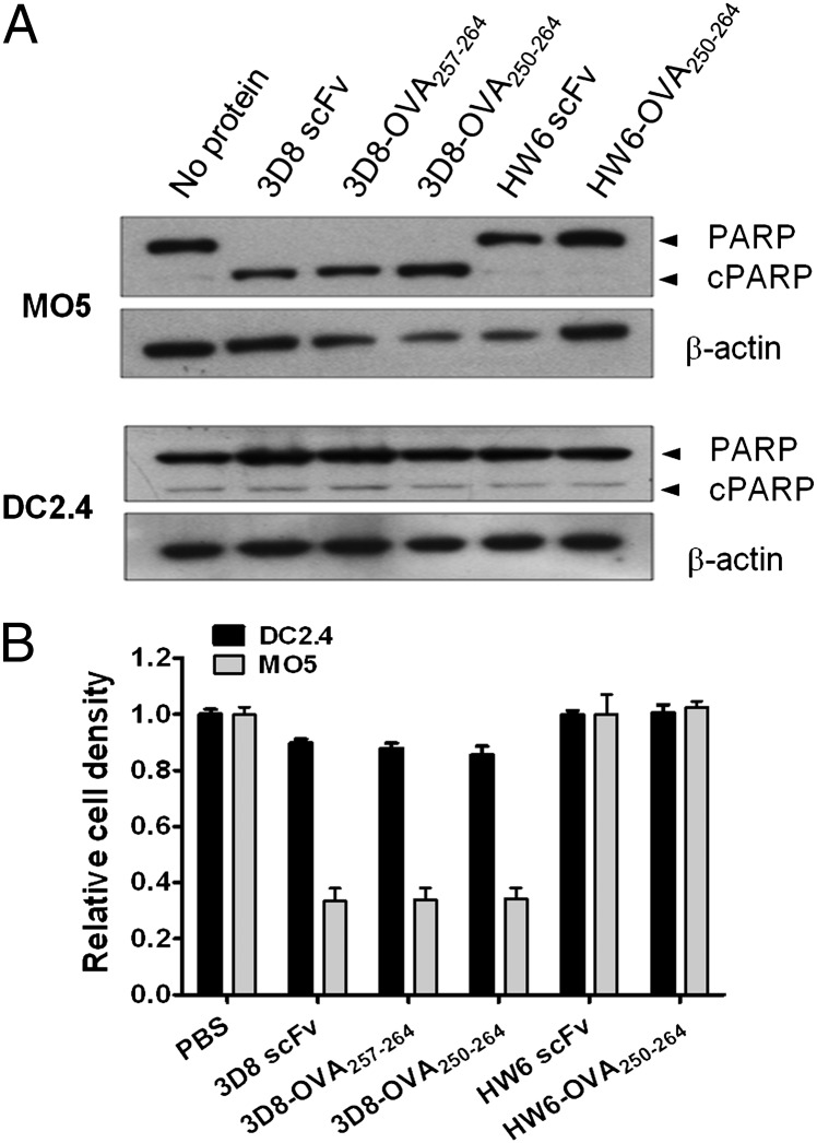
FIGURE 2.
The effect of 3D8-OVA fusion proteins on MO5 and DC2.4 cell viability. MO5 and DC2.4 cells were cultured in the presence of the indicated protein (10 μM) for 48 h; PARP cleavage was analyzed by Western blotting (A), and cell viability was determined using an MTT assay (B). The relative viability was calculated as the absorbance of the sample/the absorbance of PBS. Data represent the mean ± SE of triplicate wells and are representative of three independent experiments.

