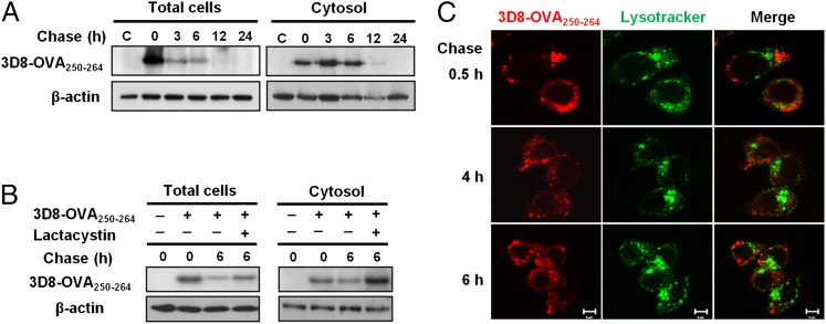
FIGURE 3.
Cytosolic translocation and proteasome-involved degradation of 3D8-OVA250–264. (A) DC2.4 cells were pulsed with 5 μM of 3D8-OVA250–264 for 2 h. Then, cells were washed and chased for the indicated times, followed by Western blotting. C, Not pulsed with 3D8-OVA250–264. Data are representative of two independent experiments. (B) DC2.4 cells were pulsed with 5 μM of 3D8-OVA250–264 in the presence or absence of lactacystin (10 μM) for 2 h followed by incubation for 6 h in the presence or absence of lactacystin. 3D8-OVA250–264 was detected by Western blotting. β-actin was used as a loading control. Data are representative of two independent experiments. (C) DC2.4 cells were pulsed with 3 μM of rhodamine-labeled-3D8-OVA250–264 (red) and then chased for the indicated times under confocal microscopy. Late endosomes and lysosomes were labeled with LysoTracker (green). Scale bar, 5 μm. Data are representative of two independent experiments.

